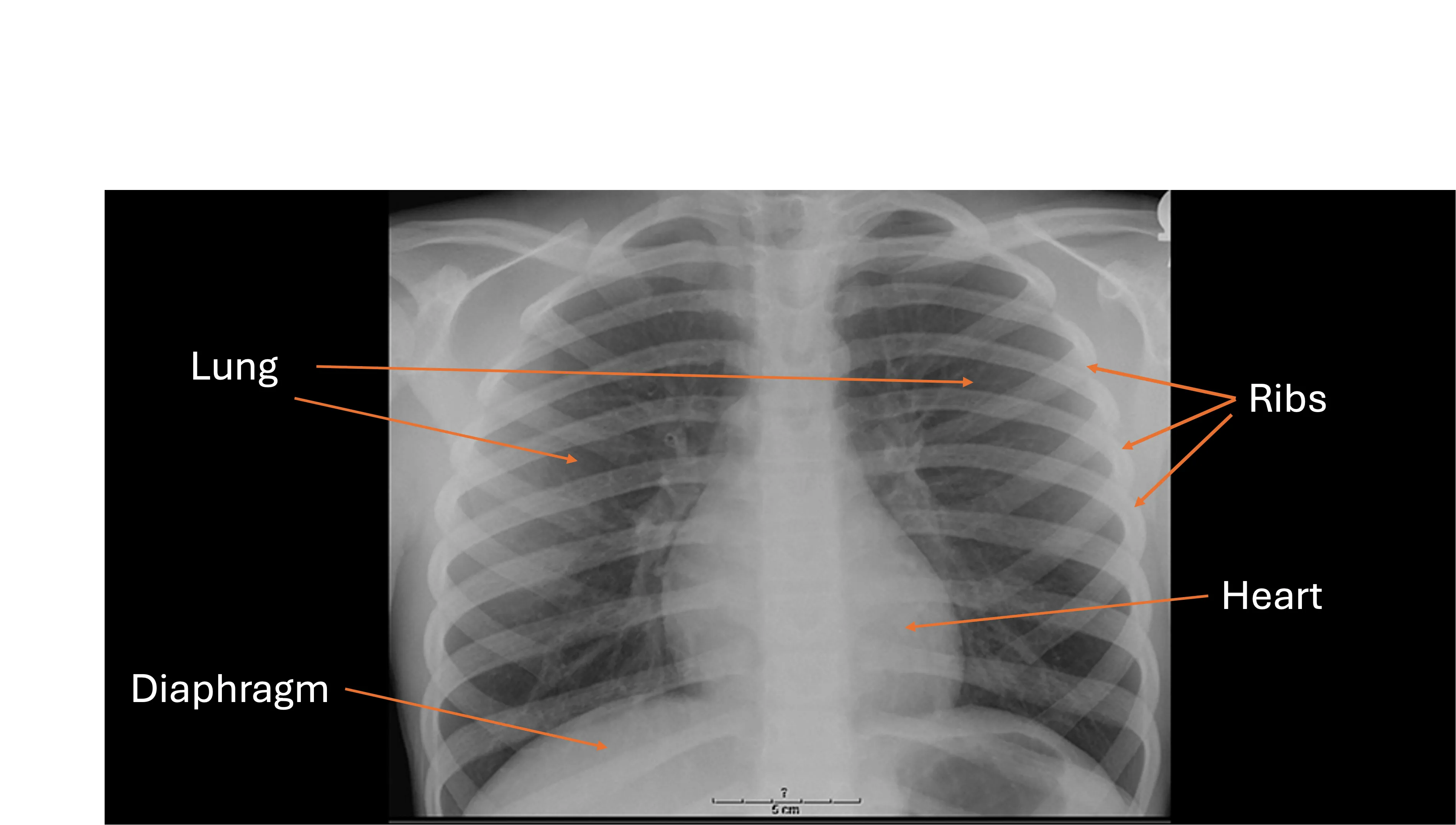
A chest x-ray creates an image of your heart, lungs, and bones (ribs and spine). Different structures allow different amounts of x-rays to pass through them. For really thick things like bones, not much passes through, so they look white on an x-ray. Your lungs allow more x-rays to pass through, so they look much darker on the x-ray.
Doctors look at the colors and shading on the x-ray to help diagnose and treat conditions.
If you get abnormal results, your doctor may recommend additional tests (like CT scans) for more information.
A chest x-ray usually involves getting 2 images — one taken from the front and one taken from the side. Your doctor will decide how many they take.
X-rays are taken by a special healthcare provider called a radiology technologist. The patient will be asked to lay down or stand next to a box that holds the x-ray equipment, and then they will turn on a light to make sure the picture is lined up correctly. Once they are in the correct position, they will be asked to hold still and take a deep breath while the technologist steps out and takes the x-ray.
It is important to hold really still during the x-ray. If your child is an infant or a toddler, they may be placed in a clear plexiglass positioner that will hold them still and in the correct position for the x-ray.
This information is for educational purposes only. It should not be used as a substitute for the medical advice of one’s healthcare provider.
